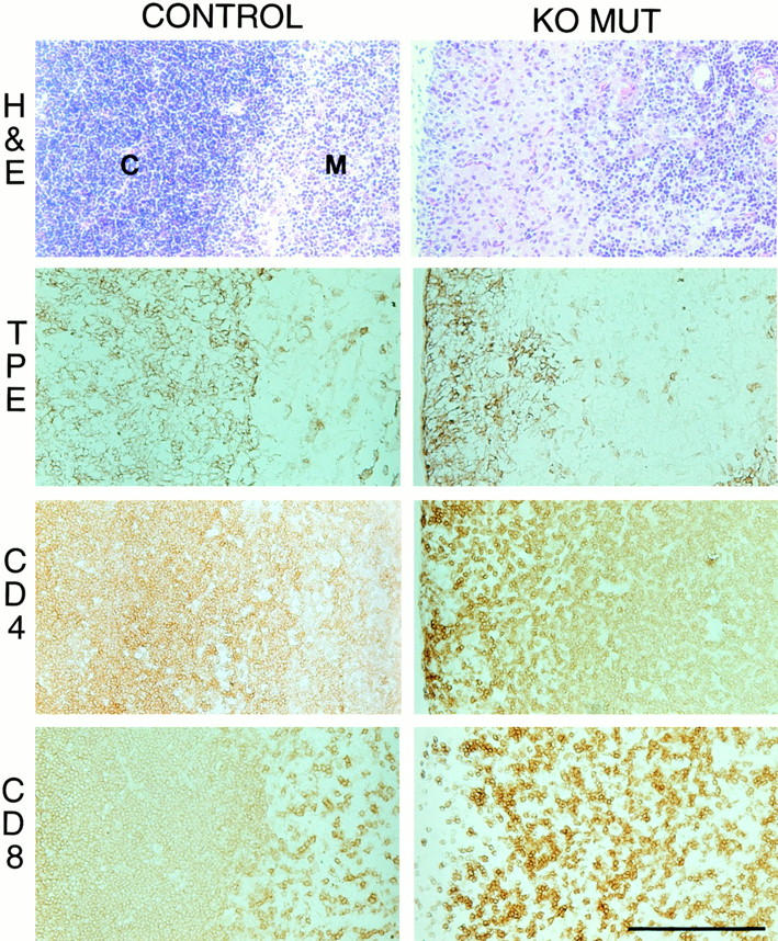
Figure 4.

Histology and immunohistochemistry of longitudinal sections of thymus showing the cortex (C)/medulla (M) junction from week three wild-type (control) and collagen X KO mice with perinatal lethality (KO MUT). In the KO MUT, H&E staining reveals reduction of cortex and a paucity of cortical T cells, whereas TPE antibodies localize the cortex as a narrow strip compared with the extensive matrix in the control. Likewise, CD4 and CD8 T cell surface markers confirm a depletion of CD4+/CD8+ immature cortical lymphocytes in the KO MUT. Bar, 100 μm.
