
Figure 2.
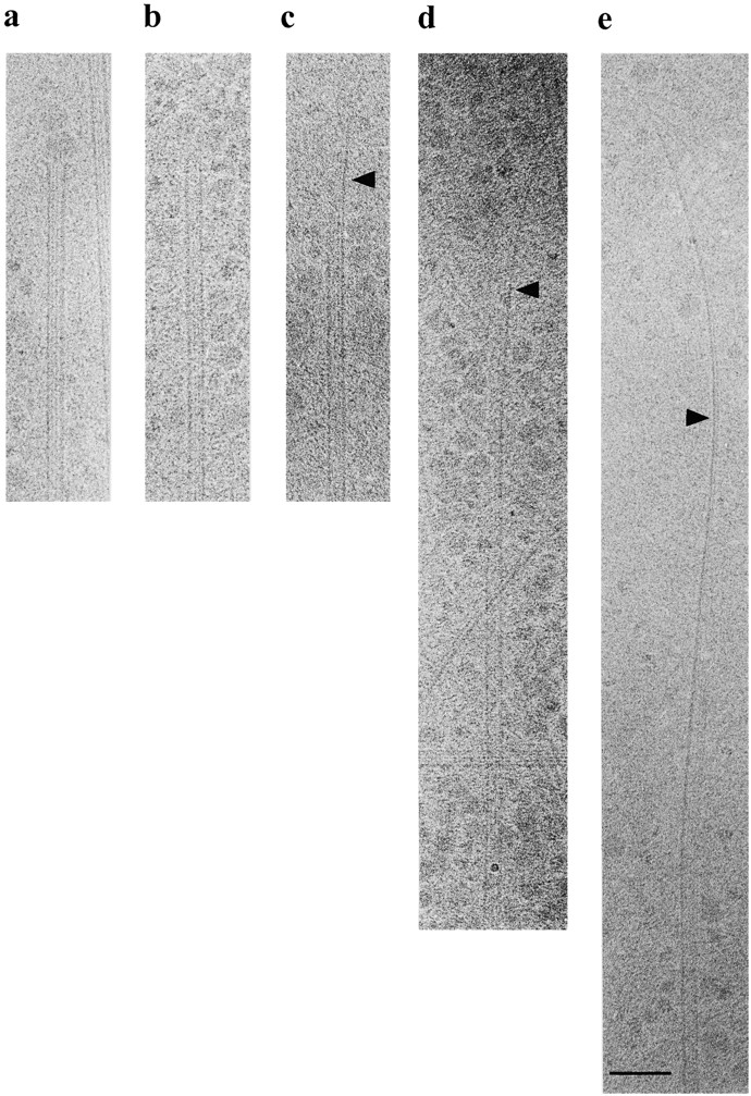
Detailed views of microtubule end structures in interphasic extracts. (a) Microtubule end with peeling protofilaments (frayed end). (b) Blunt end. (c–e) Extensions with variable lengths. Bar, 100 nm.
Official websites use .gov
A
.gov website belongs to an official
government organization in the United States.
Secure .gov websites use HTTPS
A lock (
) or https:// means you've safely
connected to the .gov website. Share sensitive
information only on official, secure websites.
Detailed views of microtubule end structures in interphasic extracts. (a) Microtubule end with peeling protofilaments (frayed end). (b) Blunt end. (c–e) Extensions with variable lengths. Bar, 100 nm.