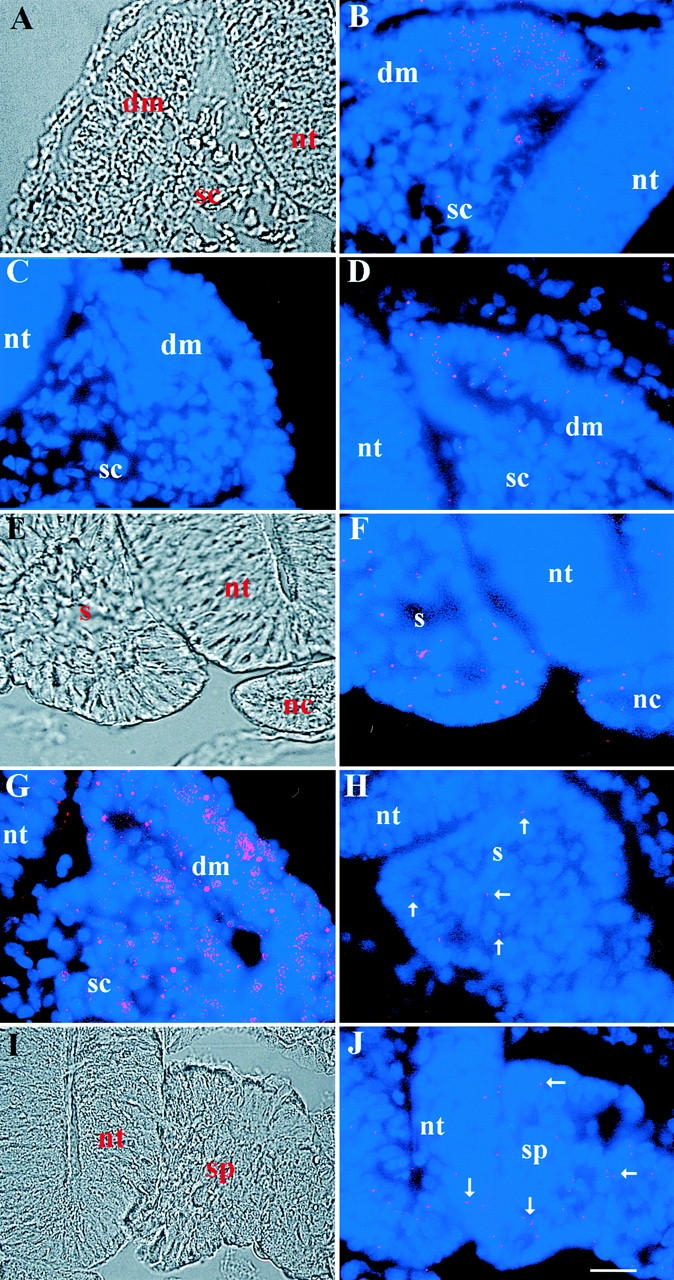
Figure 3.

Localization of MyoD and myosin mRNAs in the somites and segmental plate mesoderm of the stage 14 embryo. Photomicrographs in A, E, and I are DIC images of the merged images of bis-benzamide-labeled nuclei and Cy3-labeled dendrimers in B, F, and J, respectively. MyoD dendrimers were concentrated in the dorsal–medial portion of the dermomyotome (dm; B). A few cells of the sclerotome (sc) and neural tube (nt) also were labeled. The pattern of labeling with myosin dendrimers was similar to, but less abundant than, MyoD (D). GAPDH dendrimers produced intense fluorescence throughout the dermomyotome and sclerotome (G), whereas dendrimers lacking a specific recognition sequence did not bind to the somite (C). A subpopulation of cells in the epithelial somite (s; F) and segmental plate (sp; J) contained MyoD dendrimers. A few myosin dendrimers were found in the segmental plate (H). Bar, 9 μm.
