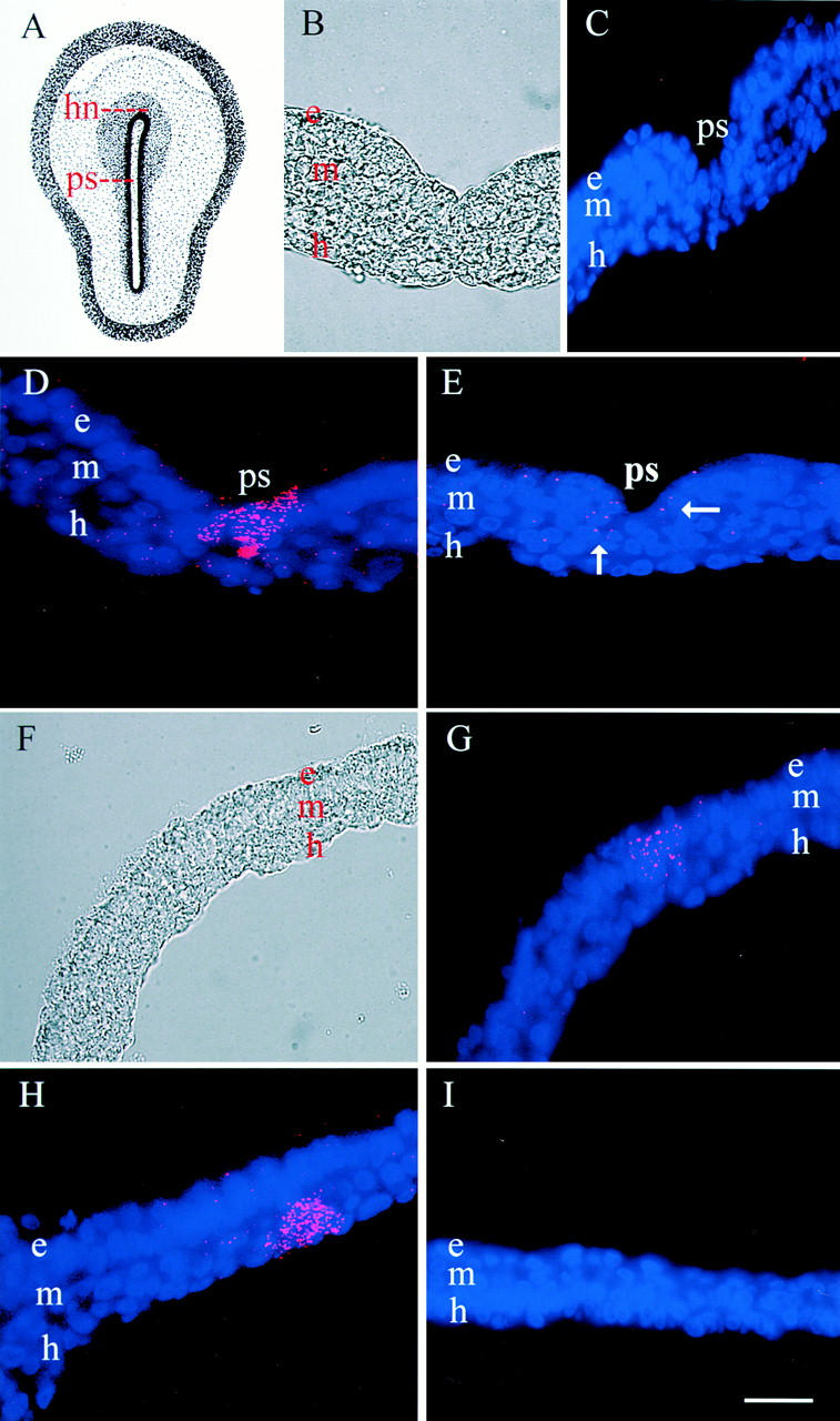
Figure 4.

Localization of MyoD in the stage 4 embryo. A drawing of the stage 4 embryo is shown in A. A DIC image of the primitive streak near Hensen's node is shown in B and the lateral region of the embryo in F. Cells ingressing into the primitive streak (ps) in the region of Hensen's node (hn) were intensely labeled with MyoD dendrimers (D). Less fluorescence was observed in more posterior regions of the streak (E). Groups of adjacent epiblast (e) and mesoderm cells (m; G) and mesoderm and hypoblast cells (h; H) also were labeled. Myosin dendrimers were not observed in the primitive streak near Hensen's node (C) or in more lateral regions of the embryo (I). Bar, 9 μm.
