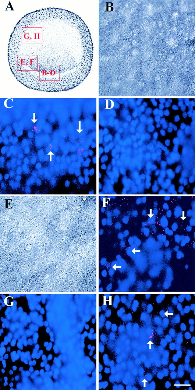
Figure 6.

Localization of MyoD in pregastrulating embryos. The areas outlined in the drawing of the pregastrulating embryo (A) are shown in B–H. Photomicrographs in B and E are the DIC images of the merged images of bis-benzamide-labeled nuclei and Cy3-labeled dendrimers in C and F, respectively. The stage X embryo contained ∼20 MyoD positive cells in the posterior epiblast (C). MyoD fluorescence extended more laterally in the posterior epiblast in the stage XII embryo (F). By stage 2, MyoD positive cells also were present in the lateral epiblast (H). Myosin dendrimers did not label stages X (D) or XII (G) embryos. Bar, 15 μm.
