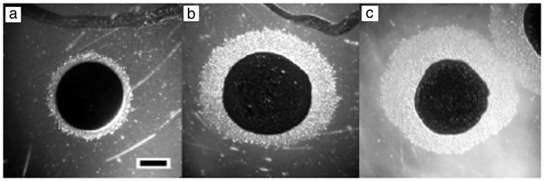
Figure 1.
M. xanthus directed movement on PE concentration gradients. Test compounds originate at the top of each panel. Cell suspensions in 10% India ink (Higgins) were spotted about 3 mm from the test compounds. (a) Delivery solvent only. (b) One microgram of PE purified from vegetative M. xanthus cells. (c) PE (0.1 μg) purified from developing M. xanthus cells. (Bar: 1 mm.)

