Figure 2.
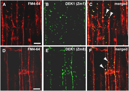
Colocalization of DEK1 and FM4-64 in Endocytotic Compartments.
(A) to (C) Confocal microscopy images of maize root cells stained with FM4-64 (A) and with the DEK1 Zm1 antibody and a secondary antibody conjugated with FITC (B). (C) shows a merged image of (A) and (B). Yellow indicates overlapping fluorescent signal (arrowheads).
(D) to (F) Confocal microscopy images of root cells stained with the endocytic marker FM4-64 (D) and DEK1 Zm8 antibody with a FITC-conjugated secondary antibody (E). (F) shows a merged image of (D) and (E). Yellow indicates overlapping fluorescent signal (arrowheads).
Note the colocalization between DEK1 antibodies and FM4-64 in punctate cytoplasmic structures, likely endosomes (arrowheads) Bars = 5 μm.

