Figure 4.
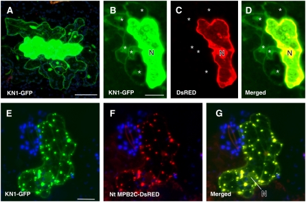
Cell-to-Cell Movement of Transiently Expressed KN1 Is Impaired by MPB2C in Arabidopsis Leaf Epidermal Tissue.
Two-week-old Arabidopsis C24 plants were bombarded with gold particles coated with plasmids expressing fluorescent proteins driven by the CaMV 35S promoter. Confocal images of fluorescent fusion proteins coexpressed in single epidermal cells were taken at 1 to 2 d after bombardment.
(A) KN1-GFP–emitted fluorescent signal (green) is present in cells adjacent to the bombarded cell at 2 d after bombardment.
(B) to (D) Coexpression of KN1-GFP and DsRED. KN1-GFP is found in neighboring cells (B), whereas DsRED remains in the expressing cell (C). (D) shows a merged image.
(E) to (G) Cell coexpressing KN1-GFP and Nt MPB2C-DsRED. KN1-GFP remains in the bombarded cell and appears in stationary punctae (E). Red channel image shows the presence of Nt MPB2C-DsRED in the same cell (F). In the merged image (G), colocalization of KN1 and Nt MPB2C fluorescent fusion proteins is visible (yellow).
N, nucleus. Stars indicate cells in which fluorescent proteins moved. Bars = 80 μm.

