Figure 1.
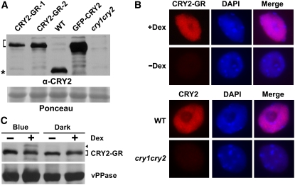
Expression and Dex-Dependent Nuclear Localization of CRY2-GR.
(A) Immunoblot showing relative levels of expression of CRY2-GR in two independent lines (CRY2-GR-1 and CRY2-GR-2), the control GFP-CRY2 in the respective transgenic lines, and endogenous CRY2 in wild-type plants. Samples were prepared from etiolated seedlings of indicated genotypes, fractioned by a 10% SDS-PAGE gel, blotted, stained with Ponceau S (Ponceau), and probed with the anti-CRY2 antibody (α-CRY2). The bracket indicates CRY2 fusion proteins, and the asterisk indicates the endogenous CRY2.
(B) Immunostaining (red) showing accumulation of CRY2-GR (line CRY2-GR-1) in the nucleus (4′,6-diamidino-2-phenylindole stain, blue) in the presence of Dex. Top panel: nuclei were isolated from CRY2-GR/cry1 cry2 seedlings grown in Murashige and Skoog (MS) medium containing 30 μM Dex (+Dex) or mock solution (−Dex) in the dark. Bottom panel: CRY2 immunostaining of the nuclei isolated from wild-type and cry1 cry2 mutant plants. The immunostaining was probed with anti-CRY2 (α-CRY2) antibody and then the second antibody conjugated with the fluorescent dye rhodamine red-x.
(C) Immunoblot comparing the level of CRY2-GR (line CRY2-GR-1) in 7-d-old seedlings grown in MS medium containing 30 μM Dex (+Dex) or mock solution (−Dex) in the dark or continuous blue light. The blot was probed with anti-CRY2, stripped, and reprobed with anti-vPPase (vacuolar pyrophosphatase) to show the relative loading. The arrowhead indicates phosphorylated CRY2-GR, and the bracket indicates unphosphorylated CRY2-GR.

