Figure 6.
Blue Light–Dependent Phosphorylation and Degradation of CRY2-GR Occur in the Nucleus.
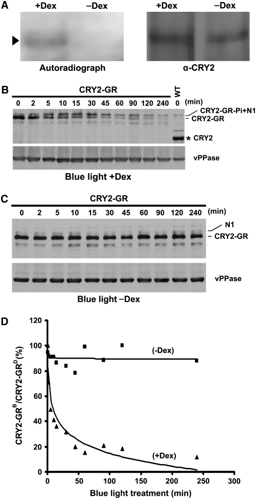
(A) Autoradiograph showing CRY2-GR was phosphorylated only in the presence of Dex. CRY2-GR/cry1 cry2 seedlings were grown on MS medium with (+Dex) or without (−Dex) Dex, preincubated with 32P for 2 h, and exposed to blue light (18 μmol m−2 s−1) for 15 min. CRY2 was isolated by IP, fractionated, and blotted. After autoradiography (left), the blot was probed with anti-CRY2 antibody (right) to show the level of CRY2. Arrowhead indicates phosphorylated CRY2-GR.
(B) and (C) Immunoblots showing that CRY2-GR underwent blue light–dependent degradation only in the presence of Dex. Five-day-old etiolated CRY2-GR/cry1 cry2 seedlings were treated with Dex (B) or mock solution (C) for 2 h and then exposed to blue light (15 μmol m−2 s−1) for the durations indicated. Protein extracts were fractioned by 10% SDS-PAGE gels, and the immunoblots were probed using anti-CRY2 and then anti-vPPase antibodies. CRY2-GR and CRY2-GR-Pi indicate unphosphorylated or phosphorylated CRY2-GR, respectively, N1 indicates the position of a band nonspecifically recognized by the anti-CRY2 antibody, and the asterisk indicates endogenous CRY2.
(D) The CRY2-GR signals were digitized and calculated as described (see Methods) and presented as the percentage of CRY2-GRB (level of CRY2-GR after blue light treatment)/CRY2-GRD (level of CRY2-GR before blue light treatment).

