
Figure 1.
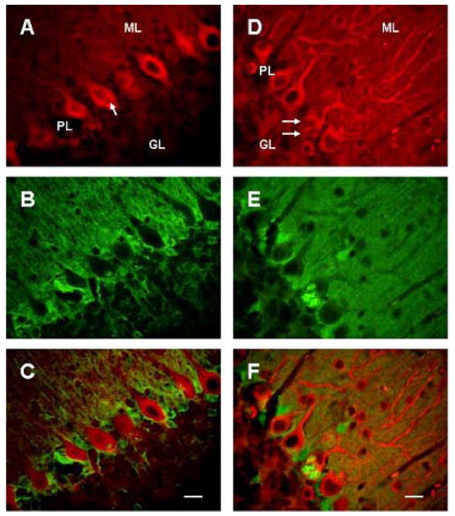
Cytoplasmic vacuoles (arrows) in Purkinje cells in 23 days (A–C) and 5 wks old (D–F) SCA1 heterozygous mice. A–C: Cerebellar section showing double immunostaining of PKC gamma (A) and S100B (B), and staining in merged image (C); bar = 10 μm. D–F: Double immunofluorscence of beta III tubulin (D) and S100B (E), and fluorescence in merged image (F); bar = 10 μm. GL, ML and PL are granule, molecular and Purkinje cell layers respectively. Vacuoles are positive for S100B and appear to be negative for both PKC gamma and beta III tubulin. Most of the vacuoles contain S100B immunoreactive central core.