
Figure 3.
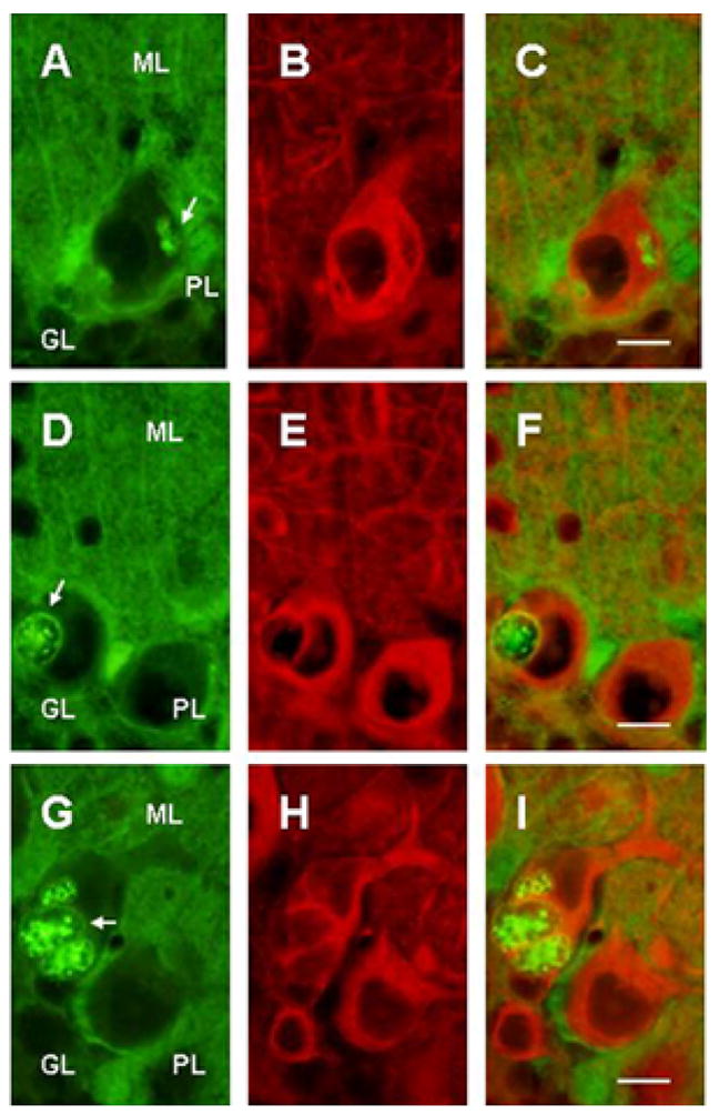
Double immunostained cerebellar sections of 5 wks SCA1 heterozygous mice showing S100B (A,D,G) and beta III tubulin (B,E,H) immunoreactions. C, F and I are merged images. The arrows indicate multiple S100B immunoreactive vacuoles of various shapes and sizes in Purkinje cells. Smaller vacuoles have somewhat homogeneous distribution of S100B protein. However, majority of vacuoles are devoid of beta III tubulin, especially in the central core. Bar = 10 μm.