
Figure 4.
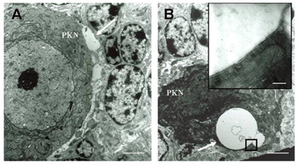
Electronmicrographs of 6wks old SCA1 heterozygous mouse cerebellum (A and B). A: A vacuole like structure, which is not yet internalized is present (arrow) near Purkinje cell’s (PKN) soma; bar = 3 μm. B: A dying Purkinje cell with two cytoplasmic vacuoles (arrow); bar = 3 μm. The inset shows that the vacuole has a double membrane; bar = 0.2 μm.