
Figure 6.
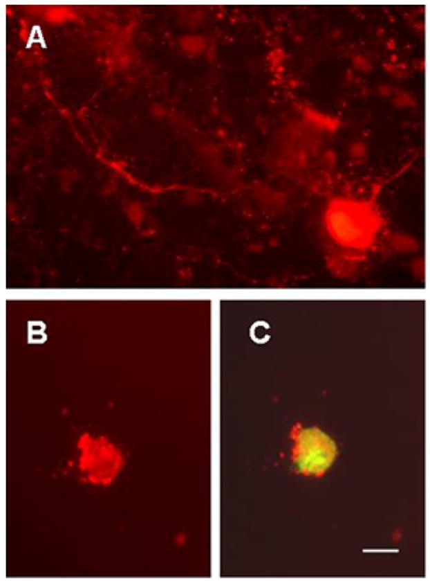
Cultured Purkinje cells showing calbindin D28k immunoreactivity (A and B) and S100B fluorescence (C) in 5 (B and C) and 12 days old (A) cultures. Purkinje cell enriched cultures were prepared from the cerebella of 0–1 day old wildtype (A) and SCA1 homozygous mouse pups (B and C). Purkinje cells were identified by size, asymmetric arbors, immunoreactivity to calbindin-D28k and failure to express glial fibrillary acidic protein (not shown). In this preparation, cultures were incubated with Oregon Green tagged S100B protein (5μg/ml) for 2 hr. Calbindin positive Purkinje cell shows internalized S100B; bar = 10 μm.