
Figure 2.
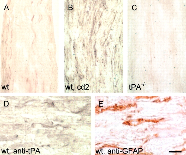
tPA is produced by Schwann cells after sciatic nerve crush. Immunocytochemistry with an antibody against tPA on longitudinal cryostat sections of wild-type, uninjured sciatic nerve revealed tPA staining only of the sciatic nerve vasculature (A). As early as 2 d after crush (cd2) tPA immunoreactivity was increased in the endoneurium (B). Sciatic nerve from a tPA−/− mouse showed no immunoreactivity with the tPA antibody (C). Immunostaining of parallel sections of a wild-type sciatic nerve 2 d after crush showed similar morphology between tPA immunoreactive cells (D) and GFAP-positive Schwann cells (E). The number of GFAP-positive cells was equivalent in wild-type and tPA−/− nerves before and after crush, suggesting that the number of Schwann cells is not significantly different in the two genotypes. Bar: (A–C) 93 μm; (D–E) 18 μm.