
Figure 3.
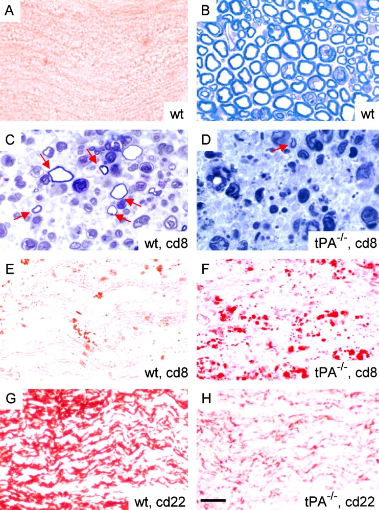
Axonal degeneration and demyelination are exacerbated in tPA-deficient mice after sciatic nerve crush. Oil Red O staining of cryostat sections of uninjured wild-type sciatic nerve (A) revealed normal myelin distribution, and toluidine blue staining of sciatic nerve semi-thin cross-sections showed normal axon morphology (B). 8 d after crush (cd8), toluidine blue staining of semi-thin cross-sections of wild-type mice (C) demonstrated more myelinated axons (arrows) than tPA−/− mice (D). Oil Red O staining showed increased accumulation of myelin and lipid debris in the tPA−/− (F) compared with the wild-type (E) sciatic nerve. 22 d after crush (cd22), staining for myelin basic protein revealed fewer myelinated axons in tPA−/− (H) than in wild-type (G) nerves. Bar, 18 μm.