Figure 1.

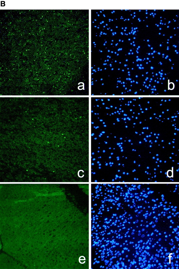
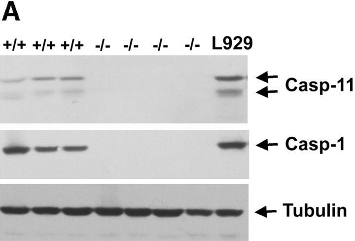
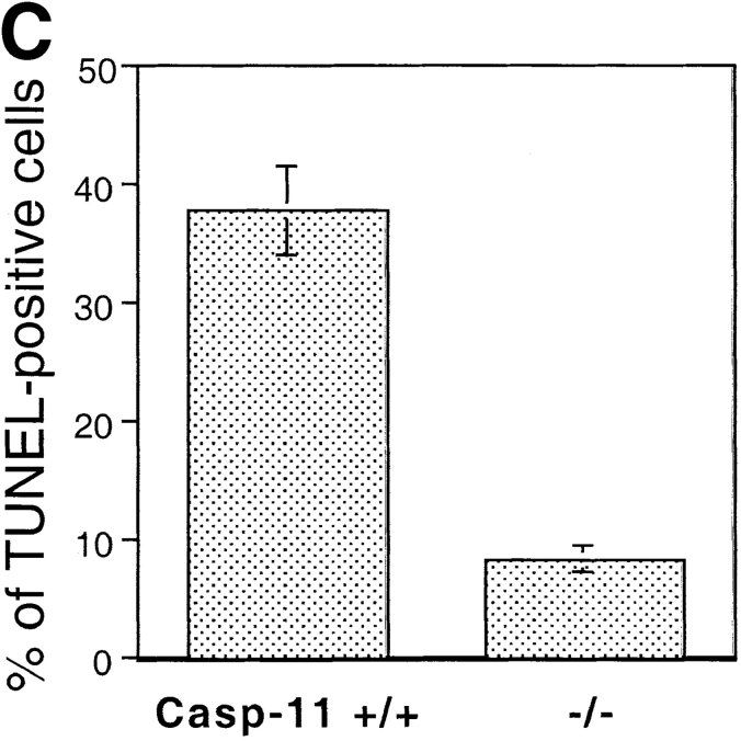
Reduction of apoptotic cell numbers after ischemic brain injury in caspase-11−/− mice. A, Western blot of spleen lysate from 3 wild-type (+/+) and 4 caspase-1 (−/−) mice, probed by anticaspase-11 (top), anticaspase-1 (middle) and antitubulin (bottom). L929 cell lysate (L929) was used as a positive control. B, TUNEL (a, c, and e) and Hoechst dye (b, d, and f) staining of wild-type (a, b, e, and f) and caspase-11−/− (c and d) brains. a–d are from ischemic brains and e and f are from untreated control brain. Permanent ischemia was induced by occlusion of the left middle cerebral artery using monofilament as described by Hara et al. 1997a. 12 h after the occlusion, brains were processed for TUNEL. C, Quantification of TUNEL-positive cells in the wild-type and caspase-11−/− ischemic brain. The percentages of TUNEL-positive cells were determined by the number of TUNEL-positive cells divided by the number of Hoechst dye-positive cells. TUNEL- and Hoechst dye-positive cells were quantified using the Zeiss Axiovert microscope equipped with Northern exposure software. Mean of the reading from five brains is shown. Error bar represents standard error.


