Figure 5.
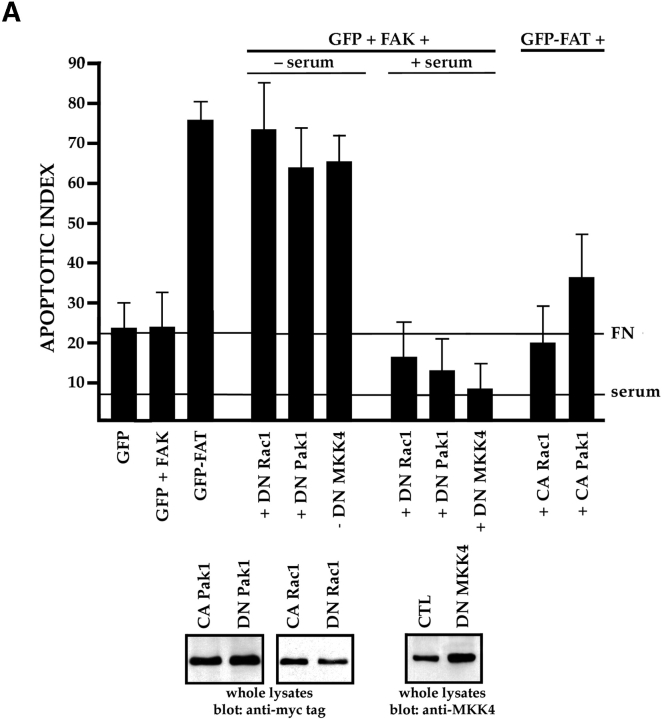
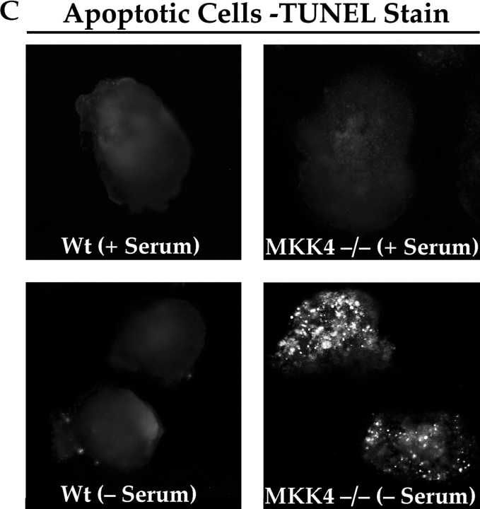
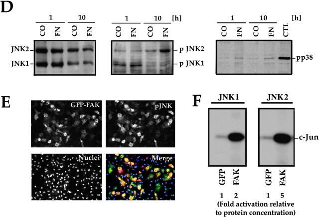
The JNK MAPK pathway transduces survival signals from FN-FAK via Rac1/Pak1/MKK4 after serum withdrawal. (A) Expression of DN Rac1, DN Pak1, or DN MKK4, along with GFP+FAK, triggered high levels of apoptosis in RSF cultured on FN in the absence, but not in the presence of serum. Coexpression of constitutively active forms of Rac1 (CA Rac1) or Pak1 (CA Pak1) rescued cells from GFP-FAT–mediated death. The Western blot panels show that CA and DN Rac1/Pak1 construct pairs were expressed at similar levels in RSF and that DN MKK4 was expressed at a level similar to the other constructs. (B) Active MKK4 was detected in lysates of cells plated on FN but not on collagen I. Lysates of RSF cultured in the absence of serum for either 1 or 10 h on FN or on collagen I were immunoblotted using antibodies that detect total MKK4 protein or pMKK4. MKK4 protein levels were similar to one another on both matrices at each timepoint. Significant levels of pMMK4 were detected only in lysates of RSF cultured on FN at the 1 h timepoint. (C) MKK4-null and wild-type (Wt) ES cells were grown in suspension for 12 d in the presence of serum. Serum was withdrawn for 24 h and embryoid bodies were processed to detect DNA fragmentation by TUNEL in situ staining. Only MKK4-null embryoid bodies showed increased TUNEL staining when serum was withdrawn. (D) JNK, but not p38 MAPK, was activated in response to attachment to FN. Lysates of RSF cultured without serum for 1 or 10 h on FN or collagen I (CO), were immunoblotted using antibodies that detect p38 MAPK protein or pp38, or JNK1/2 proteins or pJNK1/2. p38 MAPK was only weakly activated on FN. Lysates of anisomycin-treated primary RSF were used as a positive control (CTL). The signal for pJNK2 was particularly strong at 10 h in cells plated on FN. JNK1 and JNK2 protein levels in cells on FN and collagen were similar to one another at each timepoint. (E) Active pJNK (rhodamine) is strongly visible in cells transfected with GFP-FAK (green), but remains at low levels in adjacent untransfected cells (see nuclei with no corresponding GFP staining in the merged picture). Cells on FN in the absence of serum were fixed and stained after 16 h. (F) FAK, but not GFP, activates JNK1/2 in RSF. Flag-tagged JNK1 or JNK2, and FAK or GFP were coexpressed in RSF for 16 h after serum withdrawal. Cell lysates were incubated with anti-Flag antibody to precipitate JNK1/2. Precipitates were tested for their kinase activities using glutathione S-transferase–c-Jun as a substrate. JNK1/2 were activated strongly by FAK, but not by GFP.


