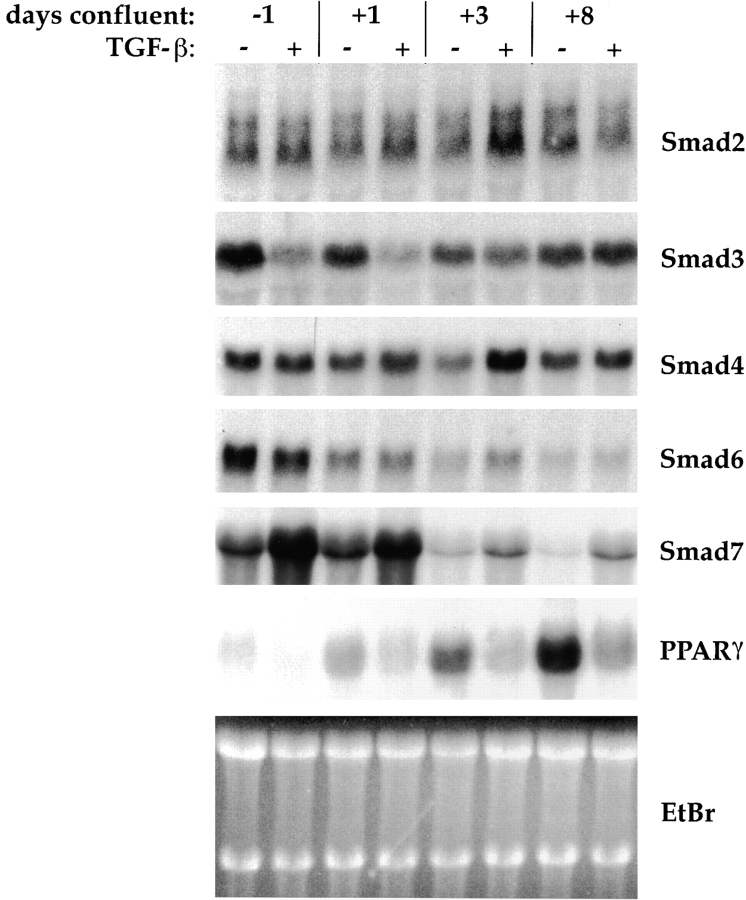
Figure 2.
Northern analysis of Smad mRNA expression during differentiation and in response to TGF-β treatment. Cells were allowed to differentiate without added TGF-β, or in the presence of 5 ng/ml TGF-β beginning at 2 d preconfluence, and RNA was harvested at the days indicated. 10 μg total RNA was blotted and sequentially hybridized to cDNA probes for Smad2, Smad3, Smad4, Smad6, and Smad7, and for PPARγ (as marker for adipogenic conversion), as indicated. Ethidium bromide staining of the gel (EtBr) is shown as a loading control. All autoradiograms were exposed for 24 h except for PPARγ, which was exposed for 8 h.

