
Figure 6.
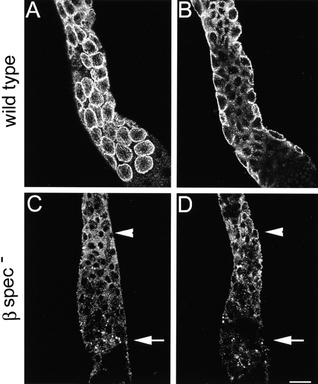
Localization of the Na,K ATPase in β spectrin mutants by confocal microscopy. Dissected preparations of the middle midgut from wild-type control larvae (A and B) and β-specem6 mutants (C and D) were stained with mouse monoclonal anti–Na,K ATPase α subunit and fluorescent secondary antibody. The basolateral staining pattern of the Na,K ATPase in wild-type appeared as rings in en face views (A), or as horseshoe shapes in optical sections near the center of the midgut (B). Neither pattern was detectable in the β-specem6 mutants (C and D). Toward the anterior (C and D, arrowhead), Na,K ATPase staining was distributed throughout the cytoplasm. Toward the posterior (C and D, arrow), staining appeared as large puncta and did not appear to be associated with the plasma membrane. In between these two regions, staining was relatively weak or in some cases absent. Bar, 10 μM.