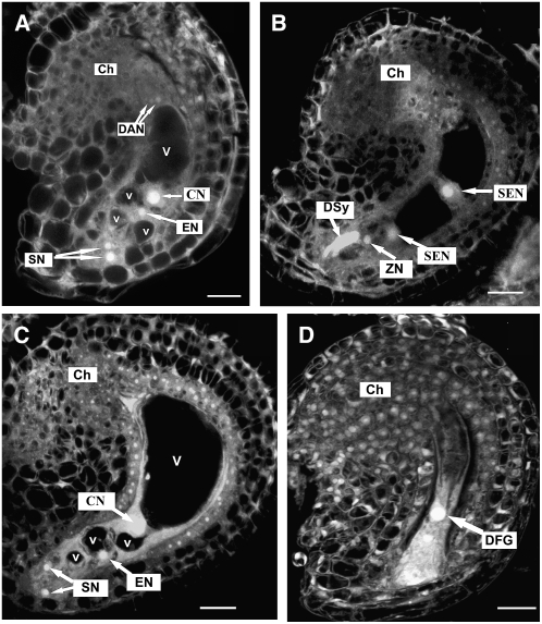
Figure 3.
Ovule Development in Mutant Plants as Revealed by CLSM Observation.
(A) A mutant ovule with mature four-celled embryo sac at early stage FG7. Note the polar nuclei fused to form a diploid central nucleus and degenerating antipodal nuclei.
(B) A wild-type ovule from a ccg/CCG pistil showing an embryo sac at 48 HAP, with one degenerated synergid cell, an intact synergid cell, a zygotic cell, and two secondary endosperm nuclei.
(C) An unfertilized mutant ovule from the same pistil as in (B) showing the four-celled embryo sac. The ovule noticeably increased in size.
(D) The unfertilized mutant embryo sac undergoes degeneration at 72 HAP. All images were projected from multiple 1-μm optical sections.
Ch, chalazal end; CN, central nucleus; DAN, degenerating antipodal cell; DFG, degenerated female gametophyte; DSy, degenerated synergid cell; EN, egg cell nucleus; SEN, secondary endosperm nucleus; SN, synergid nucleus; V, large vacuole; v, small vacuole; ZN, zygote nucleus. The developmental stages of embryo sac are defined according to Christensen et al. (1998). Bars = 10 μm.

