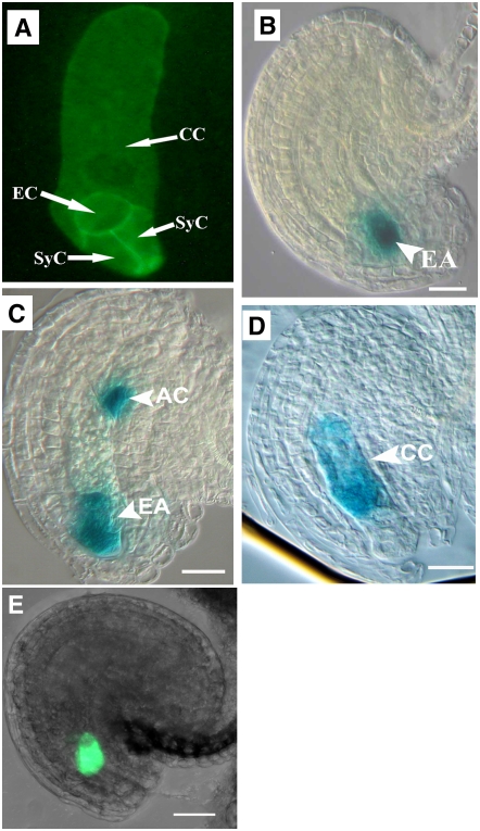
Figure 4.
Cell Fate Specification and Cellularization of Female Gametophytes in the Mutant.
Marker lines were crossed to the mutant line, and ovules were checked for marker expression pattern and statistic analysis. For ProMYB98-GFP, the reporter was introduced into the mutant by Agrobacterium tumefaciens–mediated transformation. For all five markers, the same pattern is observed in both wild-type and mutant ovules. AC, antipodal cell; CC, central cell; EA, egg apparatus; EC, egg cell; SyC, synergid cell. Bars = 20 μm.
(A) A CLSM projection showing the cellularization pattern of mature mutant embryo sacs expressing the ProAKV-GFP-ROP6C cell membrane GFP marker gene showing cellularization of the embryo sac.
(B) A mutant ovule showing egg apparatus–specific GUS marker expression (ET2632).
(C) A mutant ovule showing the expression of egg apparatus and antipodal cell marker (ET1811).
(D) A mutant ovule showing central cell–specific GUS marker expression (ET956).
(E) A CLSM image showing synergid-specific ProMYB98-GFP pattern in mutant ovules.

