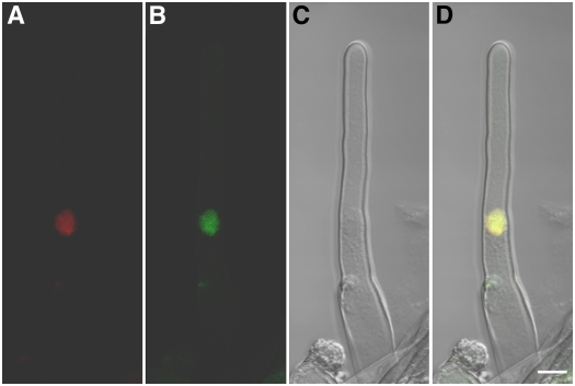
Figure 7.
Nuclear Localization of CCG Protein in Arabidopsis Transformed with the 35S∷CCG-GFP Gene.
(A) DNA 4′,6-diamidino-2-phenylindole (DAPI) staining of a root hair cell.
(B) GFP signal also detected in the same root hair cell.
(C) Bright-field image of the same cell shown in (A) and (B).
(D) A merged image of (A) to (C) showing colocalization of CCG-GFP and DAPI staining. DAPI fluorescence is coded red, and GFP signal is green. CCG-GFP localized in the nucleus. Bar = 10 μm.

