
Figure 3.
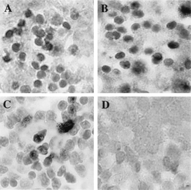
Histological analysis of VZV-infected thymus/liver implants. On day 7 after infection, thymus/liver implants infected with ROka (A), ROka47SR (B), ROka66S (C), or ROka47S (D) were fixed in paraformaldehyde, paraffin embedded, and cut into 3-μm sections before in situ hybridization was performed. A light hematoxylin counterstain was applied to reveal tissue histology. Darkly stained cells indicate VZV DNA in T cells. T cells containing VZV DNA were visible in all implants except those infected with ROka47S. (Magnification, ×786.)