
Fig. 3.
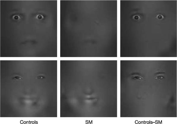
(from Adolphs et al. 2005) When exposed to isolated fragments of faces of fear and happiness, normal controls used information mostly from the eye region. In contrast, an amygdala damaged subject (SM) used information mostly form the mouth region.