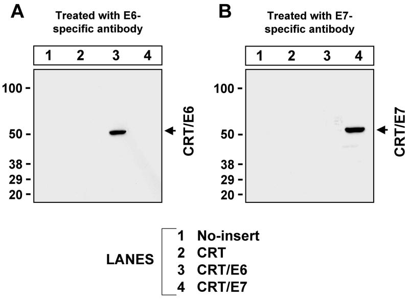
Figure 1. Western blot analysis to detect the expression of HPV-16 E6 and E7 antigens in DC-1 cells transfected with various DNA constructs.
DC-1 cells were transfected with pcDNA3-no insert, pcDNA3-CRT, pcDNA3-CRT/E6 or pcDNA3-CRT/E7. Western blot analysis was performed with 50μg of cell lysates 24 hours after transfection. E6 and E7 were detected using rabbit anti-HPV16-E6 or mouse anti-HPV16-E7 monoclonal antibody. (A) Detection of E6 expression. (B) Detection of E7 expression. The specific bands are indicated by arrows.

