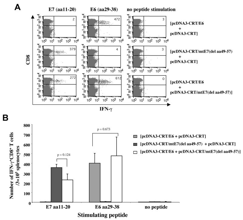
Figure 5. Intracellular cytokine staining and flow cytometry analysis of HLA-A*0201 restricted E6 and E7-specific IFN-γ-secreting CD8+ T cells in vaccinated mice.
HLA-A*0201 transgenic mice (AAD, on C57BL/6 background) (5 per group) were immunized and boosted intradermally via gene gun with the following vaccination groups: 1) pcDNA3-CRT/E6 and pcDNA3-CRT, 2) pcDNA3-CRT/mtE7(del aa49-57) and pcDNA3-CRT or 3) pcDNA3-CRT/E6 and pcDNA3-CRT/mtE7(del aa49-57). For each vaccination, the two DNA constructs were mixed evenly in the same bullet and the dose for each construct was 2μg/mouse. Splenocytes were harvested one week after the last immunization and stimulated with either E7 (aa11-20) or E6 (aa29-38) peptides. The cells were then stained for both CD8 and intracellular IFN-γ. (A) Representative figure of the flow cytometry data. (B) Bar graph depicting the number of antigen-specific IFN-gamma-secreting CD8+ T-cell precursors/3 × 105 splenocytes (mean±S.D.).

