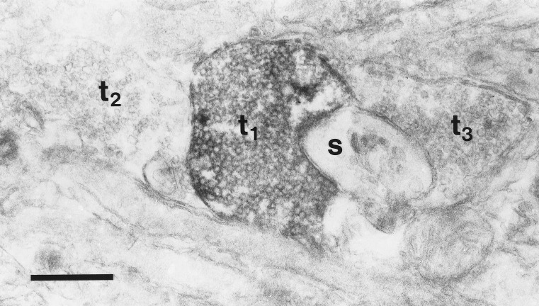
Figure 5.
Electron micrograph of phospho-p44/42 MAP kinase immunoreactivity after 30 min in EBSS without synaptic blockers. Electron-dense label can be seen in a vesicle-filled terminal (t1) in close apposition to a small, unlabeled spine (s). The flattened disk-shaped structures in the spine are characteristic of the spine apparatus. Two other unlabeled terminals are visible (t2, t3). This section was not stained with uranyl acetate or lead citrate. (Bar = 0.5 μm.)

