
Fig. 2.
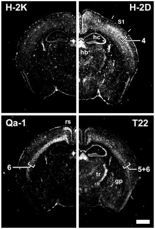
Expression of multiple class I MHC subclasses in distinct regions of the mature CNS. Coronal sections of P40 mouse brain analyzed by in situ hybridization, using subclass-specific probes indicated at top of each panel (13). S1, somatosensory cortex; hb, habenula; hc, hippocampus; rs, retrosplenial cortex; tr, thalamic reticular nucleus; gp, globus pallidus. Numerals (4, 6, 5+6) indicate neocortical layers. Scale bar, 1 mm.