
Fig. 3.
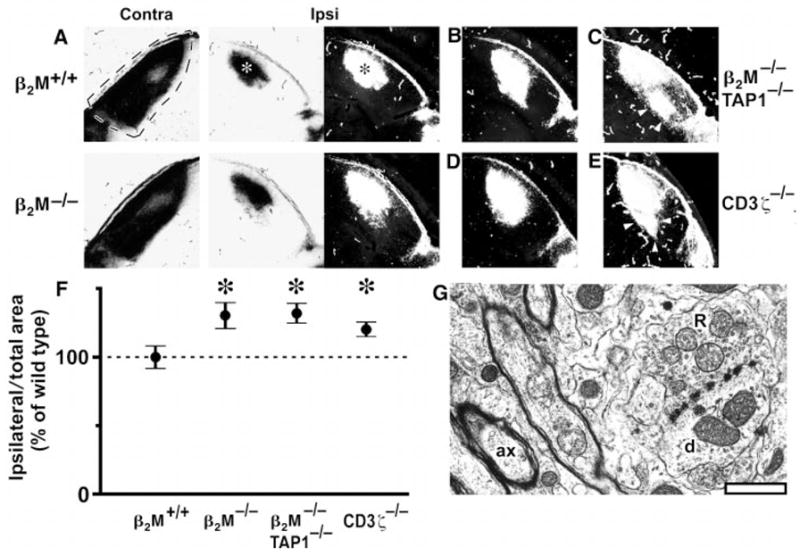
Abnormal retinogeniculate projections but normal dLGN ultrastructure in mice deficient in class I MHC signaling. At P12, one eye was injected with WGA-HRP (23); after 1 day, anterograde axonal transport results in labeling of the entire retinal projection to the LGN. Labeling pattern in the dLGN is shown in bright-field optics (label is black) or as dark-field composites [label is white; see (24)]. (A) Representative projection from retina to dLGN contralateral (dashed lines; coronal section; dorsal is up; lateral is left) or ipsilateral to eye injected with WGA-HRP (asterisks indicate labeled area from ipsilateral eye: lateral is to right) in a P13 β2M+/+ wild-type mouse and a β2M−/− mutant mouse. (B and C) Representative (B) and extreme (C) examples of the projection from the ipsilateral eye observed in β2M−/−TAP1−/− mice. (D and E) Representative (D) and extreme (E) examples of the projection in CD3ζ −/− mice. Arrowheads indicate ectopic projections, which appear extensive under the more sensitive dark-field optics. Scale bar, 200 μm. (F) Graph of areas (±SEM) occupied by the ipsilateral retinal projection to the LGN for β2M+/+ (wild-type), β2M−/−, β2M −/−TAP1−/−, and CD3ζ−/− mice (24), normalized to total dLGN area. The ipsilateral projection area in β2M+/+ animals is set as 100% (horizontal dashed line). Asterisks indicate significant differences from β2M+/+ mice (P < 0.05, Student’s two-tailed t test). (G), Electron micrograph of the dLGN from a β2M−/−TAP1−/− mouse (at P24), showing a typical R-type synaptic bouton (R) making contacts with a dendrite (d). A well-myelinated axon (ax) is also present in this field. Scale bar, 1 μm.