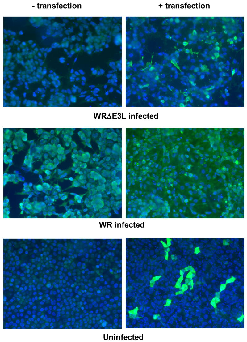
Figure 2.
Immunofluorescence (IF) showing that MAb 3015B2 is reacting specifically to VACV E3 protein. RK-13 cells seeded on an 8-chamber glass microscope slide (Nunc) were either untransfected or transfected for 24 hours with 2 μg of pMT-E3L using FuGene 6 reagent (Roche). Cells were then either left uninfected or infected with WR or WRΔE3L viruses for 24 hours. Cells were then fixed with 4% formalin, permeabilized with 0.1% Triton X-100 in PBS, and then probed with hybridoma generated mouse ascites at a 1:200 dilution. Cells were then washed with 3% bovine serum albumin (3% BSA-PBS), probed with goat anti-mouse immunoglobulin G conjugated to fluorescein isothiocyanate (FITC) (Zymed) at a 1:200 dilution, and washed with PBS. The nuclei of the cells were stained with DAPI (Vector Labs). Cells were visualized using a Nikon Eclipse E1000 fluorescence microscope with the appropriate filters and analyzed using Phase 3 Imaging software.

