
Figure 5.
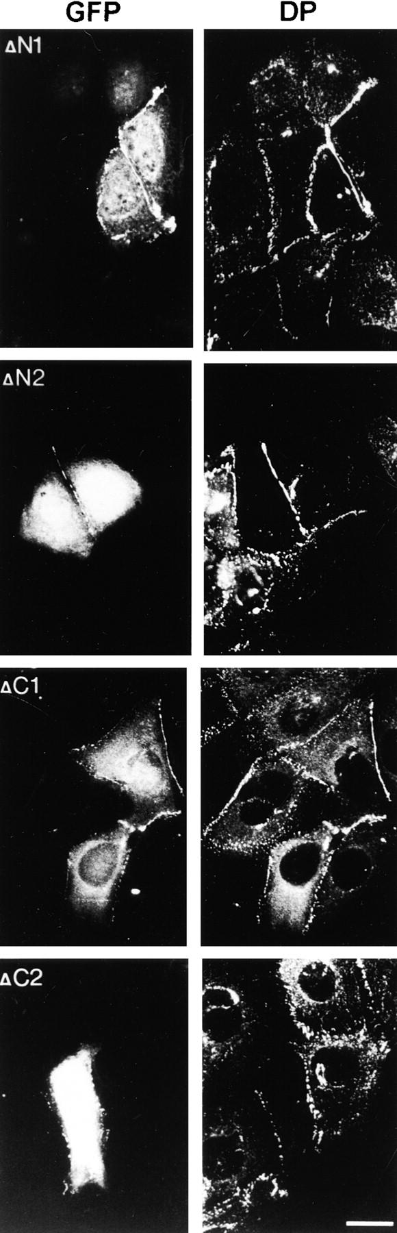
Expression of plakophilin 1 head domain fragments in HaCaT cells. Plasmid DNAs encoding the GFP-tagged plakophilin 1 head domain fragments were transfected into HaCaT cells, and their ability to recruit desmoplakin to the cell membrane was analyzed by immunofluorescence. Whereas desmosome localization was found with all fragments, nuclear staining was strong only with ΔN2 and ΔC2. ΔN1 and ΔC1 showed reduced nuclear staining. Desmoplakin recruitment to the plasma membrane, as revealed by continuous labeling along the cell periphery, was enhanced with ΔN1, ΔN2, and ΔC1. In ΔC2-overexpressing cells, desmoplakin staining showed no considerable increase. Bar, 20 μm.