
Figure 6.
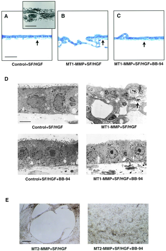
Invasion of peritoneal tissue by MDCK transfectants in vivo. A, Vector control-transfected MDCK cells cultured on the peritoneal matrix (arrow) did not express invasive activity after stimulation with SF/HGF for 12 d in complete media. Bar, 100 μm. The inset shows the peritoneal matrix by TEM after the mesothelial cell layers were removed. The tissue contains numerous collagen and elastin fibrils sandwiched between two discontinuous basement membranes. Bar, 1 μm. B, MT1-MMP–overexpressing cells degraded large blocks of tissue and invaded into the underlying collagen after stimulation with SF/HGF for 12 d. A small segment of remaining peritoneal tissue is indicated by the arrow. C, In the presence of BB-94, MT1-MMP transfectants failed to invade the peritoneum after 12 d in culture (arrow). D, TEM analysis of peritoneal tissue invasion by control or MT1-MMP transfectants cultured for 12 d with SF/HGF in the absence or presence of BB-94 as indicated. Only a portion of the peritoneum can be identified after culture with MT1-MMP–overexpressing cells (arrow). Bar, 1 μm. E, MT2-MMP–overexpressing cells completely degraded entire tracts of the peritoneum after stimulation with SF/HGF for 12 d as shown in an en face view of a live culture. The addition of BB-94 to these cultures blocked tissue dissolution and maintained an intact peritoneal matrix. Bar, 100 μm.