
Figure 1.
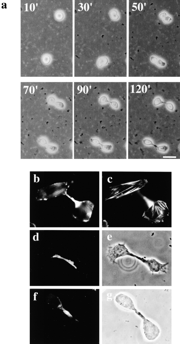
SA-Ro treatment blocks disassembly of the contractile ring. a, Time-lapse observation of cytokinesis of cells treated with SA-Ro. The time shown in the photograph represents time points from the initiation of cell division (min). Bar, 30 μm. b and c, CHO cells synchronized in prometaphase were incubated with 50 μg/ml SA-Ro for 2 h, and then fixed and stained with rhodamine-phalloidin. b, Shows the focal plane of the cytoplasmic bridge where actin bundles remains concentrated. c, Shows the focal plane of the bottom layer of the daughter cells where stress fibers are seen. d–g, Distribution of myosin II (d) and radixin (f) in SA-Ro–treated cells. Phase-contrast of each specimen was shown in e and g, respectively. Bar, 10 μm.