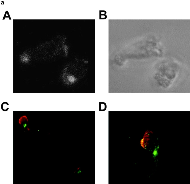
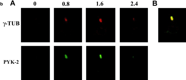
Figure 1.
PYK-2 is localized at the uropod projection of NK cells adhered to fibronectin. (a) Confocal section of PYK-2 immune staining of NK cells migrating on FN (A). The same section under brightfield illumination is shown in B. Epifluorescence images of NK cells double-stained for PYK-2 (green) and moesin (red, C), and PYK-2 (green) and talin (red, D). Control cells stained with goat serum or secondary antibody alone did not show any specific staining, and preincubation of the anti-PYK-2 antiserum with its specific blocking peptide prevented staining of NK cells (not shown). (b, A) Confocal serial sections of polarized NK cells adhered to FN and double-stained for γ-tubulin (red, upper panels, γ-TUB) and PYK-2 (green, lower panels, PYK-2). Sections were taken every 0.8 μm from the substratum (0) in the z-axis. b, B shows a merged image of double-labeled cells demonstrating the colocalization of γ-tubulin and PYK-2 at the uropod of NK cells.


