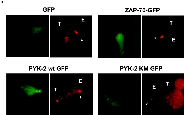
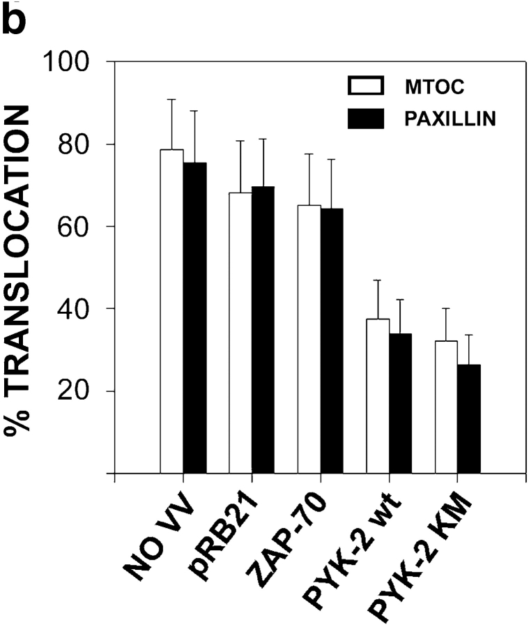
Figure 8.
Overexpression of PYK-2 wt-EGFP and PYK-2 (K-M)-EGFP prevents the translocation of MTOC and paxillin during specific target recognition. (a) NK inhibitory clones were infected with control VV-pRB21-EGFP (EGFP) (upper left panels), ZAP-70-EGFP (upper right panels), PYK-2 wt-EGFP (lower left panels), and PYK-2 (K-M)-EGFP (lower right panels). NK cell conjugates with the sensitive .221 target were studied by immunofluorescence. EGFP green fluorescence shows the infected NK cells (left panels). The red fluorescence corresponds to γ-tubulin (MTOC) staining (right panels). T and E indicate target and effector cells, respectively, while arrowheads point to the MTOC on effector cells. (b) Quantification of the translocation of MTOC and paxillin in conjugates formed by a green (infected) NK cell from inhibitory clones and its sensitive target .221. Translocation of MTOC or paxillin was measured in >100 conjugates in three independent experiments. Results correspond to the arithmetic mean ± SD.


