
Figure 6.
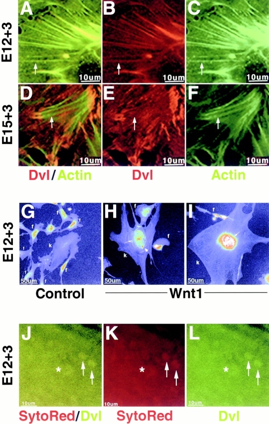
The subcellular localization of dishevelled is regulated during nephric tubule induction and by Wnt1 signaling. (A–F) Embryonic kidney cells were harvested at E12 (A–C) or at E15 (D–F) and cultivated for 3 d on collagen, followed by fixation in 100% cold methanol and immunofluorescence staining using anti-dvl and antiactin antibodies. (A–C) Cells from E12 kidneys exhibit a colocalization of endogenous dvl along actin stress fibers (arrows). (D–F) Cells from E15 kidneys exhibit decreased colocalization of dvl with actin stress fibers (arrows) and, instead display a punctate pattern of dvl staining on the basal cell surface and an accumulation of dvl at the end of actin stress fibers. (G–I) Wnt1 stimulation leads to the accumulation of dvl in and around the nucleus of metanephric cells. E12 kidney cells (k) were cultured for 2 d on collagen-coated coverslips and cocultured with control or Wnt1-expressing NIH 3T3 fibroblasts (f) for an additional 24 h. Cells were fixed in 0.5% glutaraldehyde and endogenous dvl was detected using anti-dvl antibodies. Pseudocolor was used to quantify the amount of nuclear dvl and to visualize cell boundaries. Control-treated metanephric cells possessed low levels of nuclear dishevelled (G), whereas Wnt1-treated metanephric cells exhibited high levels of nuclear and perinuclear dvl staining (H and I). In particular, dvl accumulated between nuclei (H and I, thin arrow) and in a punctate pattern adjacent to the nucleus (H, thick arrow). Wnt1 treatment also increased the incidence of polynucleated metanephric cells and promoted cell spreading (H and I). (J–L) Dvl is present in the nuclei of metanephric mesenchymal cells in E12 kidneys cultured ex vivo for 3 d on collagen-coated coverslips and fixed in 0.5% glutaraldehyde. The presence of dvl inside nuclei was determined by observing colocalization of dvl immunofluorescence and the nuclear stain SytoRed signal in confocal optical sections (arrows). Nuclear dvl (arrows) was observed in cells on the cortex of the developing kidney proximal to the tips of ureteric bud branches (asterisk), where mesenchyme is induced to transform into tubular epithelium.