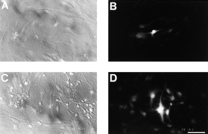
Figure 1.
Dye coupling between astrocytes is upregulated by neurons. Dye coupling was determined by patch clamp recordings of astrocytes performed with a pipette solution containing 0.2% LY. A–D, Light micrographs taken with Hoffman optics of (A) astrocyte culture and (C) spontaneous coculture and fluorescence micrographs of the same fields (B and D, respectively) taken 5 min after withdrawal of the recording pipettes. The number of dye-coupled cells is increased in the presence of neurons, indicating an increase of the permeability of gap junctions. Bar, 100 μm. Bottom, Summary diagram of dye coupling experiments obtained from 184 astrocytes filled with LY and classified in three categories, noncoupled (0), weakly coupled (1–10; stained cells), and highly coupled (>10 stained cells). The threeway χ2 comparison test for the three categories reveals a significant difference in the distribution of astrocytic coupling measured in the absence and presence of neurons (P < 0.01).


