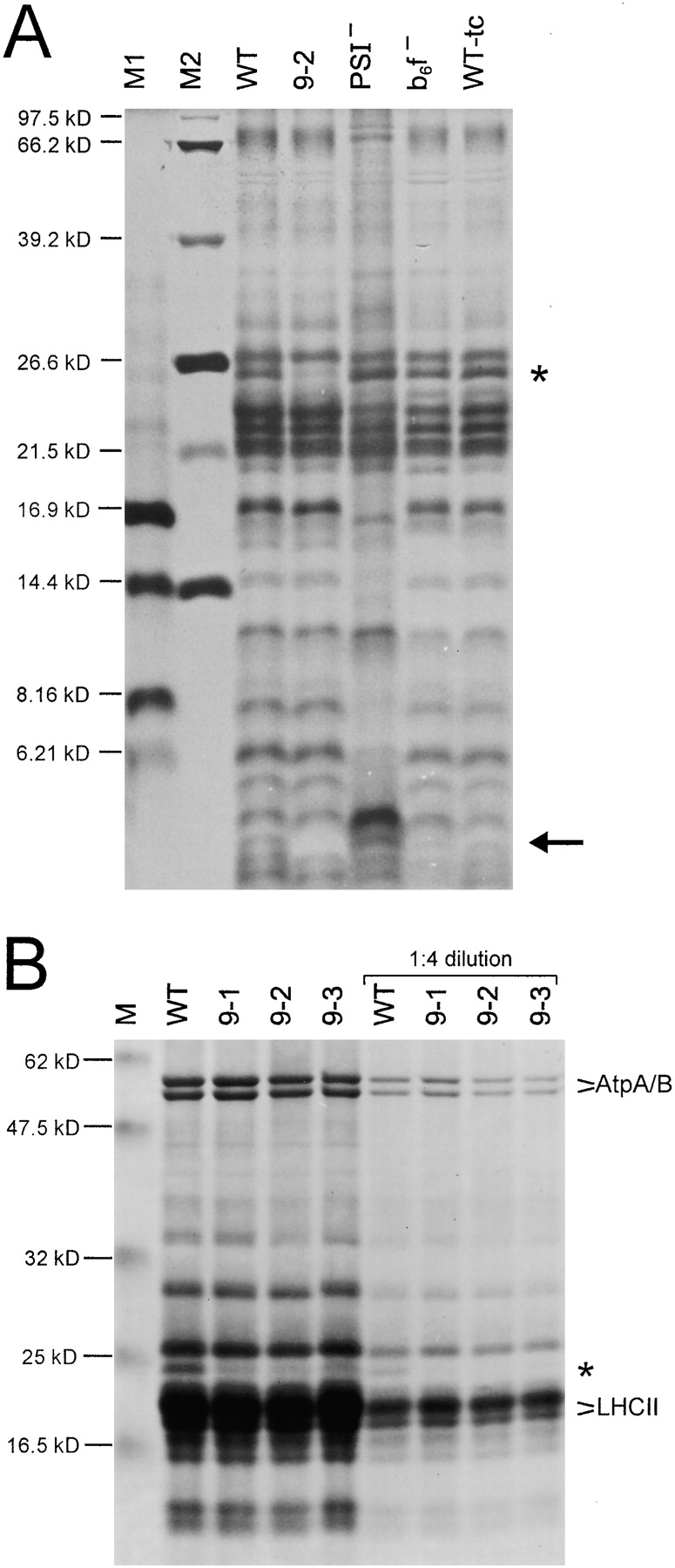
Figure 4.

Analysis of thylakoid proteins from wild-type (WT) and ycf9 knockout plants. A, Silver staining of thylakoid proteins separated by electrophoresis in a 16.5% polyacrylamide (PAA) gel. For comparison, a mutant lacking PSI (PSI−; Ruf et al. 1997), a mutant lacking the cytochrome b6f complex (b6f−; Hager et al. 1999) and a wild-type in tissue culture (WT-tc) are also shown. The putative Ycf9 protein is marked by an arrow and is clearly present in all samples except for the thylakoids from the ycf9 knockout (9-2). Note that, as expected for a hydrophobic protein, the putative Ycf9 protein migrates faster than a protein of the calculated molecular weight of Ycf9 (6.58 kD). The asterisk marks a protein specifically reduced in ycf9 knockout plants (see part B). M1, M2: molecular weight markers. (B) Coomassie staining of thylakoid proteins from the wild-type and three independently generated ycf9 knockout plants (9-1, 9-2, and 9-3). Thylakoid proteins were electrophoresed in a 10% PAA gel. Two different protein concentrations were loaded. All ycf9 knockout plants clearly show specifically a drastic reduction of a thylakoid protein with an approximate molecular weight of 25 kD (asterisk). Selected prominent thylakoid proteins are marked (AtpA/AtpB, LHCII proteins). M, molecular weight marker.
