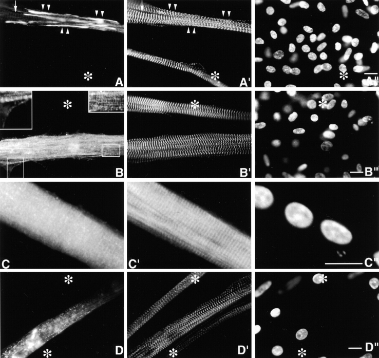
Figure 7.
Day 10 MYC/M185-SH3–transfected culture, triple stained with (A) anti-MYC, (A′), anti–s-α-actinin and (A′′) DAPI. Ectopic MYC-positive fibrils/patches (arrowheads) are negative for s-α-actinin. They do not act as dominant negatives. Arrows point to just perceptible normal Z-bands. Day 10 MYC/M185-Ser–transfected culture stained with anti-MYC (B and C), anti–s-α-actinin (B′), Rho-phalloidin (C′), and DAPI (B′′ and C′′). The ill-defined MYC-positive striations shown in B (insets) does not perturb the normal s-α-actinin Z-bands shown in B′. Similarly, the diffuse MYC/granules in C does not alter the multiple Rho-phalloidin cross-bands (C′). Day 10 GFP/M86-M92–transfected myotube viewed in the fluorescein channel (D), in the rhodamine channel (D′) after decoration with rhodamine-labeled anti–s-α-actinin, and in the DAPI (D′′). Similar images were observed in MYC/M86-M92–transfected myotubes (not shown). The GFP-positive fibrils/patches evident in D do not deform the s-α-actinin Z-bands shown in D′. Asterisks mark untransfected myotube in Fig. 7. Bars, 10 μm.

