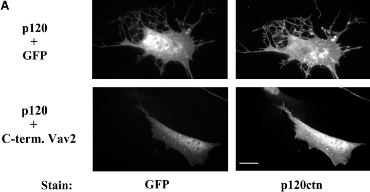
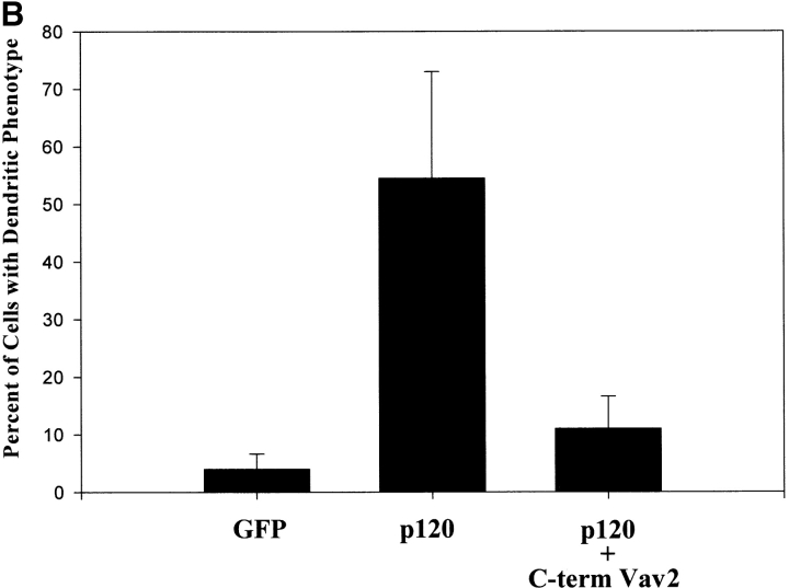
Figure 9.
The p120ctn-induced dendritic phenotype is suppressed by coexpression of the COOH terminus of Vav2. NIH3T3 cells were plated on coverslips and transfected with p120ctn + GFP (top panel) or p120ctn + COOH terminus of Vav2 fused to GFP (C-term Vav2) (bottom). Left panels show GFP fluorescence of cells transfected with GFP or C-term Vav2-GFP and right panels are stained with anti-p120ctn antibodies. (B) Transfected cells were scored for the dendritic phenotype (see Fig. 2 C) and compared. Data are the means ± SD of four separate experiments.


