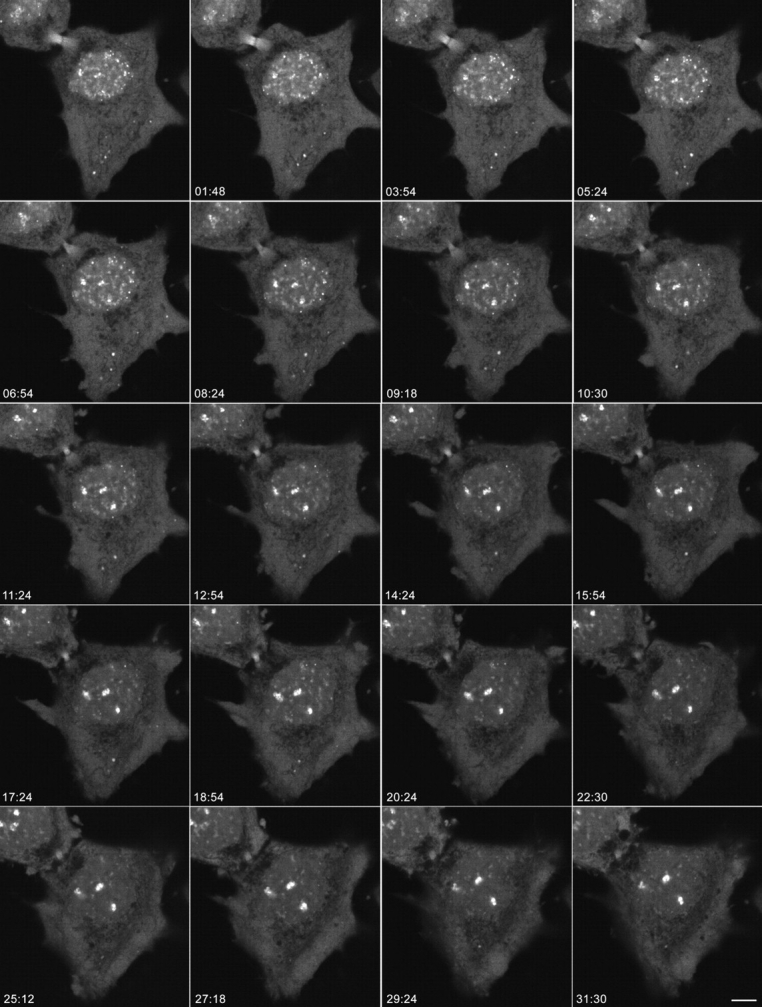
Figure 3.

Dynamics of nucleolar reassembly in telophase. CMT3 cells in telophase, which were transiently expressing the fibrillarin-GFP protein, were subjected to time-lapse confocal fluorescence microscopy. The images were collected every 18 s over a 60-min period. See also video 2 available at http://www.jcb.org/cgi/content/full/150/3/433/DC1. The series of frames shows the progression of the cell from early to late telophase with transfer of material from PNBs to the newly forming nucleoli (the three brightest spots in the nuclei). Note the gradual disappearance of the PNBs in the nucleoplasm and the NDF in the cytoplasm with a concomitant increase in the fluorescent signal in the nucleoli. Bar, 10 μm.
