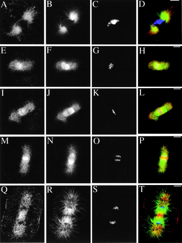
Figure 3.

Immunolocalization of KRP180 in one-cell cleavage stage sea urchin (S. purpuratus) embryos. Whole embryos were methanol-fixed and stained with anti-KRP180 (A, E, I, M, and Q) and an FITC-conjugated anti–α-tubulin antibody (B, F, J, N, and R). Anti-KRP180 was recognized with Cy5-conjugated secondary antibody and DNA was visualized with (1 μg/ml) DAPI (C, G, K, O, and S). Merged images are shown with KRP180 in red, tubulin in green, and DNA in blue (D, H, L, P, and T). Prophase (A–D), prometaphase (E–H), metaphase (I–L), anaphase (M–P), and telophase/cytokinesis (Q–T). Bar, 10 μm.
