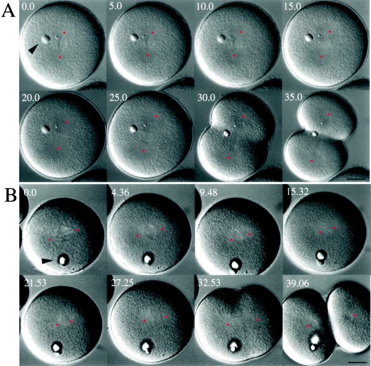
Figure 6.

Effects of anti-KRP180 and control antibody microinjected into mitotic sea urchin (L. pictus) embryos. Video frames of representative control IgG (A) and anti-KRP180 (B) injected embryos are shown. The video frames (with time shown as minutes and seconds) show the effects of microinjection on first cleavage in both embryos during NEBD (time 0.0 min) until the completion of cytokinesis (time 35.0 min for control and 39.06 for experimental embryo). Both embryos were microinjected in interphase, shortly after fertilization. The center of each pole is marked with a red dot to assist in measuring spindle pole-to-pole distances. The intracellular oil droplets (arrowheads) confirm that both embryos were microinjected successfully. Bar, 20 μm.
