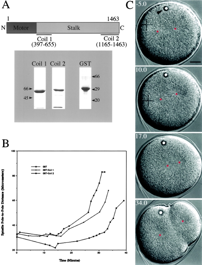
Figure 8.

Effects of GST-KRP180 dominant/negative protein microinjected into mitotic sea urchin (L. pictus) embryos. (A) Purified GST-KRP180 stalk fusion proteins (Coil 1 and Coil 2) were used as dominant/negative proteins. The numbers below the linear map of the KRP180 polypeptide correspond to the amino acids that comprise the fusion proteins. Molecular mass markers indicate the size of the glutathione–Sepharose-purified fusion proteins (lane Coil 1 and 2; Coomassie-stained 7.5% and lane GST; 12% SDS-PAGE). (B) Microinjection of GST-Coil 2 into mitotic embryos perturbs the maintenance of prometaphase spindle shape, whereas microinjection of GST-Coil 1 and control GST does not. Spindle pole-to-pole distances were measured over time for individual spindles in GST-, Coil 1–, and Coil 2–injected embryos. Time 0 represents NEBD and the final time point indicates the completion of cytokinesis. Anaphase B initiates ∼15 min post-NEBD in the three spindles shown. (C) Video frames of a representative GST-Coil 2 injected embryo are shown. The video frames (with time in minutes) show the effects of microinjection on first cleavage five minutes before NEBD (time −5.0) until near the completion of cytokinesis (time 34.0). This embryo was microinjected in interphase, shortly after fertilization. The center of each spindle pole is marked with a red dot to assist in spindle length measurements while the intracellular oil droplet (arrowhead) indicates a successful microinjection. This embryo divides asymmetrically due to mispositioning of the spindle after displaying a prometaphase spindle collapse.
