
Figure 8.
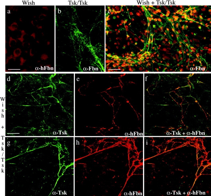
Immunofluorescence of WISH cultures using the mAb201 (α-hFbn) antibody (a), Tsk/Tsk cultures using the pAb9543 (α-Fbn) antibody (b), and WISH and Tsk/Tsk cocultures using the pAb9543 (α-Fbn) (c), pAb8368 (α-Tsk) (d and g), or mAb201 (α-hFbn) antibody (e and h). f and i show the superimposition of the images in d and e and g and h, respectively. The antibody pAb9543 (α-Fbn) was employed to better appreciate the morphology (b) and extent (c) of the microfibrillar aggregates; additionally, nuclei of cocultured cells were stained with propidium iodine to document the level of cell confluency. Bars: (a and b) 20 μm; (c) 60 μm; (d–i) 40 μm.