
Figure 9.
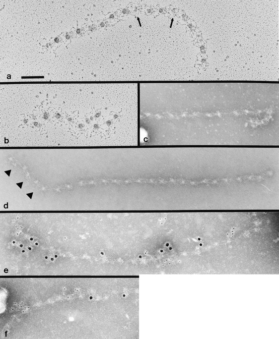
Electron microscopy of fibrillin 1 microfibrils extracted from the matrix of cocultures of WISH and Tsk/Tsk fibroblasts and viewed after rotary shadowing (a and b) or after negative staining (c–f). Arrows in a indicate abnormal globular morphologies. Arrowheads in d indicate irregularities in periodicity. Microfibrils in e and f are labeled with mAb201 (recognized by the large, 10-nm, gold conjugate), specific for human fibrillin 1, and pAb8368 (recognized by the small, 5-nm, gold conjugate), specific for mouse Tsk fibrillin 1. Bar, 100 nm.