
Figure 3.
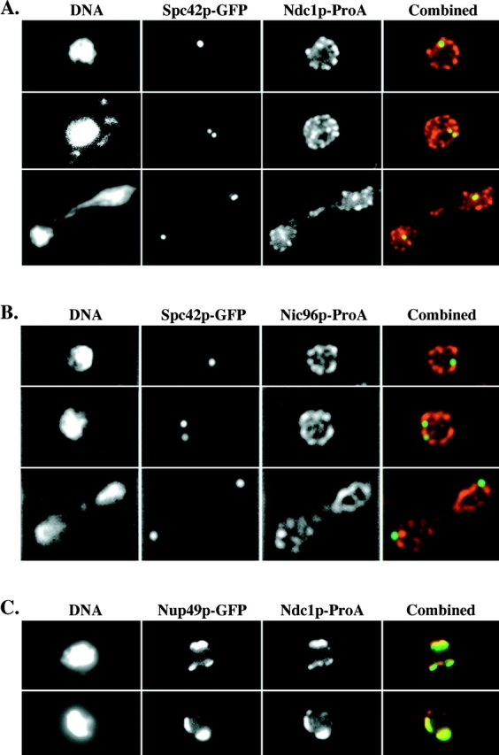
Ndc1p-ProA localizes to SPBs in whole cells. (A) Indirect IF deconvolution microscopy of cells (HC22-2b/1c; Table I) containing Ndc1p-ProA and Spc42p-GFP. Ndc1p-ProA was detected using a Texas red–conjugated secondary antibody, and Spc42p-GFP was detected by means of GFP autofluorescence using FITC filters (see Materials and Methods). The combined image shows both Texas red (red) and FITC (green) signal (overlap = yellow). (B) Indirect IF deconvolution microscopy of cells containing Nic96p-ProA and Spc42p-GFP (HC23-11d/16a; Table I) serves as a negative control for A. Samples were prepared as in A. The combined image shows both Texas red (red) and FITC signal (green). (C) Indirect IF deconvolution microscopy to examine the colocalization of Ndc1p-ProA with Nup49p-GFP in a nup133 null strain that exhibits NPC clustering (HC27-16d/16b; Table I). Ndc1p-ProA was detected as in A, and Nup49p-GFP was detected using FITC filters. The combined image shows both Texas red (red) and FITC (green) signals (overlap = yellow). Shown here are examples of cells that contain either one or two extra spots of Ndc1p-ProA staining that do not colocalize with Nup49p-GFP signal.