
Figure 1.
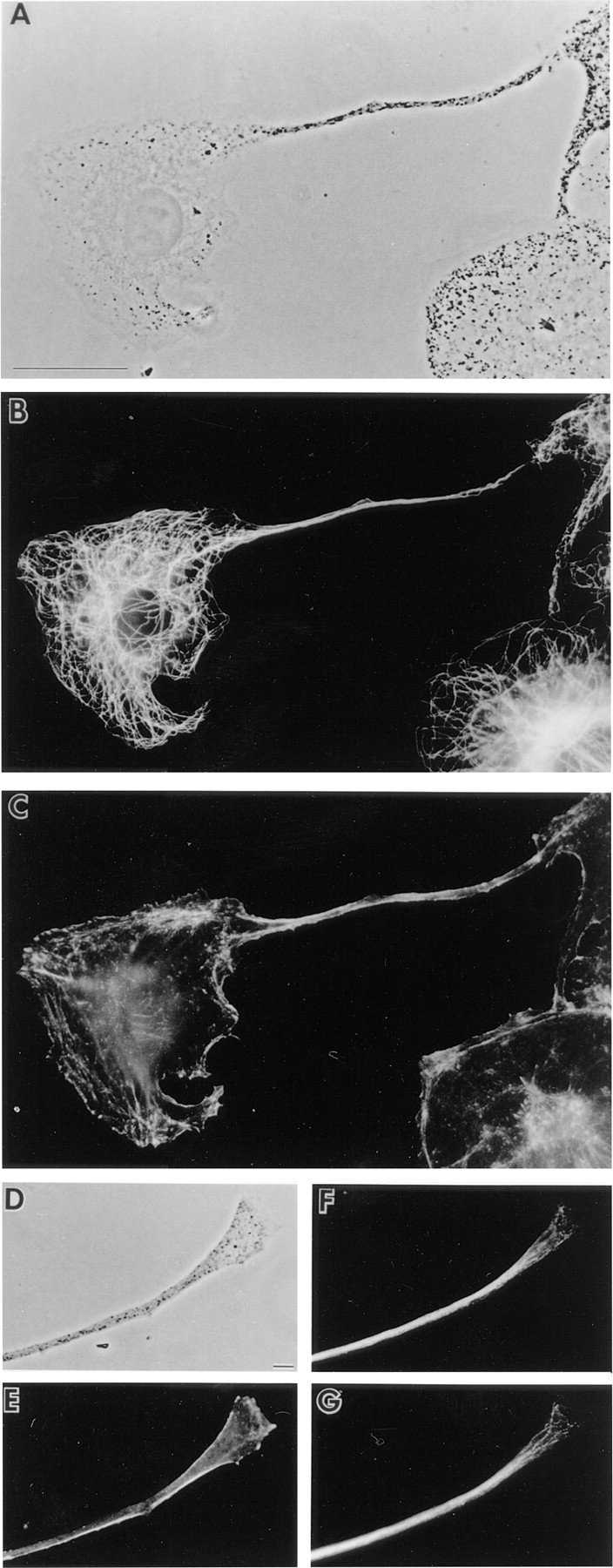
Organization of microtubules and F-actin in melanocytes. Shown are phase contrast images of a melan-a melanocyte exhibiting a single, long dendritic extension (A) and the distal portion of another dendrite (D), and the distributions of microtubules (B, F, and G; F and G are two different focal planes) and F-actin (C and E) within them. Bars: (A) 10 μm; (B) 2 μm.