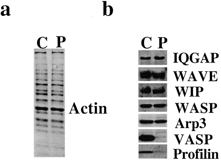
Figure 2.
Treatment of supernatant with PLP depletes profilin and VASP. (a) Coomassie blue–stained SDS-PAGE of PLP bead–treated supernatant (P) and control (Sepharose bead–treated) supernatant (C). Little or no change in protein staining was detected. (b) Western blots of PLP-treated supernatant (P) and control supernatant (C) stained with antibodies to IQGAP, WAVE, WIP, WASP, Arp3, VASP, and profilin. Of these, only VASP and profilin were decreased by PLP treatment. Each gel lane was loaded with 30 μg protein.

